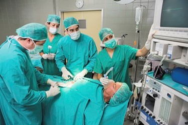
Holograms Provide Unique 3-D Views In The OR

By Katie Wike, contributing writer

Technology that converts 2-D images into 3-D images provides a unique virtual view of patients’ bodies for operating room doctors.
Surgeons will soon be able to navigate their patients’ bodies with the help of 3-D holograms. According to Newsweek, technologies like MRIs and CTs have given doctors a look inside their patients without making a single cut, but they can’t yet see these images in 3-D.
“Doctors are trying to extract 3-D information from flat slices,” says Sergio Aguirre, the founder and chief technology officer of EchoPixel, a tech company trying to solve the flat-screen problem. “We found they are looking at flat image slices, and correlating between images, and in the process, forgetting what they are looking at.”
iHealth Beat explains EchoPixel’s technology converts a series of 2-D images, such as those from a CT scan into a 3-D model. The software can be used with special 3-D glasses so that clinicians can view a projection of the model above a display board.
“It’s important to clarify that many of these new technologies aren’t real holograms, which perfectly recreate the patterns of light that reflect off an object in real life,” Wired Magazine writes. “What EchoPixel (which prefers to call their technology 3-D imaging) and others are doing is closer to stereoscopic vision, like what you see in 3-D movies or gadgets like the Nintendo 3DS.”
EchoPixel’s software is currently in use at Stanford University, the University of California-San Francisco and the Cleveland Clinic.
“All you have to do is look at how CT and MRI affected health care,” says Dr. David Langer, chief of neurosurgery at Lenox Hill Hospital. “All of a sudden we saw things we never saw before, and we started to develop new strategies for treating problems we never knew about. [3-D imaging] could result in absolutely the same thing.”
